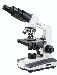
Application of biological microscope in biological research

Application of biological microscope in biological research http: //? Id = 290
Atomic force microscopy has played an irreplaceable role in biological research since its advent, and is an indispensable tool in life science research. The atomic force microscope (AFM) technology itself has many advantages, such as simple sample preparation, operation in multiple environments, high resolution, and so on. This article mainly reviews its application in biology from the aspects of biochemistry, cell biology, immunology and material ultrastructure research.
RNA
(AFM) advantages
(AFM) is a kind of 80, SPM). In 1986, Dr.
Binning won the Nobel Prize in Physics for inventing the scanning probe microscope. The magnification of this microscope is far more than any previous microscope: the magnification of optical microscopes is generally no more than 1,000 times; the magnification limit of electron microscopes is 100101000 times, which can directly observe the molecules and atoms of matter. The further exploration provides an ideal tool.
(AFM) itself is the main reason for its rapid development in biology. First of all, the sample preparation of the atomic force microscope (AFM) technology is simple and does not require special treatment of the sample. Therefore, its destructiveness is more destructive than other commonly used biological techniques (AFM) (AFM) can operate in multiple environments () Direct imaging under physiological conditions, and real-time dynamic observation of live cells; third, three-dimensional image of atomic force microscope (AFM) / submolecular resolution; fourth, atomic force microscope (AFM) can observe local areas with nanometer resolution The charge density and physical properties of AFM can manipulate individual biomolecules. In addition, the information obtained by AFM can also be complementary to other analytical techniques and microscope techniques.
(AFM) also has the ability to process the molecules or atoms of the specimen. For example, it can move atoms, cut chromosomes, and punch holes in cell membranes. In summary, the high resolution of the atomic level and the observation of the force behavior of living samples and processed samples have achieved the three characteristics of atomic force microscopy.
Principle of Atomic Force Microscope (AFM)
(AFM) usually uses silicon nitride as a sensitive elastic microcantilever, with a sharp probe at its tip for scanning on the sample. The interaction force between dots or atoms is usually described by Lennard-Jone potential: U (r) =-U0 [(r0 / Z) 12- (r0 / Z) 6] where Z, U0 and r0 are respectively The energy and distance between atoms in equilibrium. When the distance between atoms is less than r0, the force between atoms changes from attractive to repulsive. The attractive force and repulsive force between the probe and the surface are used in the scanning force test. Probe forces at different surface orientations give information about surface morphology and some other surface characteristics. Atomic force microscopy (AFM) is based on repulsion and attraction, respectively. In the atomic force microscope (AFM) test ,: F (Z) = 2Ï€R0B / 3Z3, where Z and R0 are the radius of the tip of the microsphere, B (AFM), after a laser beam emitted by the laser diode is reflected by the cantilever, hit On a split-type photodiode, because the atomic structure of the sample surface fluctuates, an atomic structure image of the unevenness of the sample surface can be obtained. Atomic-weight surface morphology records are atomic force microscopes (AFM)
Tapping mode (Tapping
Mode, TM) (AFM) is a key advancement in the observation of soft, sticky, and fragile samples. TM-AFM contacts and leaves the surface alternately at a frequency of 50,000 to 500,000 times per second. Due to the energy loss caused by the tip contacting the surface, the cantilever oscillation is weakened. This reduction in amplitude can be used to identify and measure the surface state. When the needle tip passes through the raised part of the surface, the cantilever oscillates in a smaller space, and the amplitude of the oscillation becomes smaller at the same time; on the contrary, when the needle tip passes through the depression, the amplitude becomes larger. The digital feedback loop is used
Adjust the tip-sample distance to maintain a constant amplitude and force on the sample. TM-AFM, in order to avoid the whole liquid cells into the up-and-down movement state driven by the cantilever oscillation, an appropriate oscillation frequency (usually 5000 ~ 40,000 times /) must be selected. The characteristics of this method are: when the needle tip is evacuated from the sample surface in the XZ direction and then approached, and the distance of each withdrawal is kept equal, if the needle tip is evacuated far enough, the lateral force of the needle tip on the sample will not be accumulated Can reduce the damage of the needle tip to the sample. The advantage of TM-AFM imaging is that it can not only prevent the adhesion of the needle tip to the surface and the sample damage caused by the scanning process, but also contact the surface and obtain high-resolution images. And TM-AFM imaging has a wide linear range, allowing repeated testing of conventional samples.
Observe the biochemical process using atomic force microscopy (AFM)
With the improvement of sample processing technology in liquid imaging technology, it is possible to observe complex biochemical processes using atomic force microscopy (AFM). The transcription process is the central link of gene expression, and the use of atomic force microscopy (AFM) to observe the interaction between protein and DNA has a contradiction to be resolved: biomolecules need to be fixed to the substrate is the basis of atomic force microscopy (AFM) imaging, and biochemical reaction But it requires biomolecules to move relatively freely. Even in the presence of large amounts of non-specific DNA, RNA polymerase (RNAP) and the promoter still have a high binding rate, and it is suspected that the diffusion of RNAP along the DNA is one of the reasons. After the non-specific complex is deposited under appropriate conditions, using atomic force microscopy (AFM), it can be observed that RNAP slides along the DNA and can be transferred between different DNA fragments. However, the addition of heparin can terminate these processes, which further confirms the RNAP-
Non-specific DNA interactions. Atomic force microscopy (AFM) can also observe the transcription process in real time. After adding nucleotides, the extended complex deposited on the mica moves unidirectionally along the DNA template. Two control experiments confirmed that the relative movement of RNAP and DNA is consistent with the actual situation of transcription. In a control, microcircle DNA without a terminator was used as a template for transcription on mica. After drying, the long chain of synthetic RNA can be observed by atomic force microscopy (AFM). In the second control, DNA was transcribed on mica under the same conditions. The difference is that the added nucleotide is labeled with 32P. Analysis of the reaction products by PAGE showed that the complex bound to mica is active, and the speed of transcription and the conformational changes of approximate biomolecules measured by atomic force microscopy (AFM) are also important observations of atomic force microscopy (AFM). The urease was deposited on mica and scanned with an atomic force microscope (AFM). After adding urea to the liquid pool, it was found that the vertical fluctuation of the cantilever increased significantly, which suggests that the conformational changes caused by the enzyme activity can be directly recorded by the atomic force microscope (AFM) . The outer membrane of Gram-negative bacteria is its protective barrier, which is composed of regularly assembled protein channels. Among them, the most researched is Deinococcusradiodurans' hexagonal assembly intermediate (hexago
nallypacked intermediate, HPI) protein. HPI is believed to be related to nutrient intake and metabolite excretion. HPI's Atomic Force Microscope (AFM) images show regular hexagons and a central hole-like structure. When imaging in liquid, it was found that HPI exhibits two different conformations, "on and off." Although the meaning is unclear, it shows the advantages of atomic force microscopy (AFM) imaging in liquid.
The application of atomic force microscopy (AFM) in the study of molecular recognition The interaction between molecules is quite common in the field of biology, such as the binding of receptors and ligands, the binding of antigens and antibodies, the binding of information transmission molecules, etc. The basis of information transmission in the body. Atomic force microscope (AFM) can be used as a force sensor to study the interaction between molecules. This is because the atomic force microscope (AFM) can theoretically sense a force of 10-14N and a displacement of 0.01 nm, and the contact area can be as small as 10 nm2. Therefore, atomic force microscopy (AFM) is used to study the interaction between complementary DNA strands, cell adhesion molecules, and ligand-receptor interactions. There is a high affinity between biotin and streptavidin, and the thermodynamic data of their interaction is also clear. Therefore, biotin and streptavidin are good examples for the determination of specific interaction force by atomic force microscopy (AFM). In a classic experiment, biotinylated calf serum albumin (biotinlated
The bovine serum albumin (BBSA) wraps the microspheres, and the microspheres are attached to the cantilever to form a BBSA functionalized probe. The adhesion between the BBSA functionalized probe and the BBSA-coated mica was then measured in a streptavidin solution with and without biotin block. The results show that the biotin-blocked streptavidin solution requires a large force to separate the BBSA functionalized probe from the mica surface. The force is (0.257 ± 0.025) nN, which is separated from the ligand -The force required by the receptor matches. On this basis, the effective fracture distance can be calculated as (0.95 ± 0.10) nm. Therefore, when the needle tip is wrapped with a specific molecule (such as biotin), the interaction between the needle tip and the sample can be used to identify the position of the corresponding molecule on the surface (such as streptavidin). Commercially modified probes have appeared, and these probes encapsulate different molecules and can be used for molecular recognition for different purposes. Therefore, the atomic force microscope will play a wider role.
The application of atomic force microscopy (AFM) in the study of the ultrastructure of materials can directly observe surface defects, surface reconstruction, morphology and position of surface adsorbents, and surface reconstruction caused by surface adsorbents. Atomic force microscopy (AFM) can observe the atomic-level flat structure of many different materials. For example, the atomic force microscope (AFM) can be used to study DL-leucine crystals. The ordered arrangement of surface crystal molecules can be observed, and the lattice spacing Consistent with X-ray diffraction data. In addition, atomic force microscopy (AFM) has been successfully used to observe the surface of organic molecules and biological samples such as trisorbic acid, DNA, and proteins adsorbed on the substrate. Alginate Alginate
Poly L-Lysine Alginate (APA) capsule film has semi-permeability, which can prevent the components of the human immune system from entering the capsule made of APA film, so that the contents of the capsule are protected from the immune system. Therefore, the film capsule can be used to protect the transplanted tissue in the human body and prolong its survival time in the human body. At the same time, it has a slow-release effect on the drug. The semi-permeability of APA film is closely related to the ultrastructure of its surface. It is of great significance to study the ultrastructure of its surface to study its semipermeability. There have been reports in the literature on the use of atomic force microscopy (AFM) to study the surface structure of the APA film, and the special structure of the APA surface has been discovered, thus revealing the significance of the ultrastructure of the APA surface for semi-permeability. At present, images of ultrastructures of DNA, dialysis membranes, alkane molecules, fatty acid membranes and polysaccharides have been obtained using atomic force microscopy (AFM).
Application of Atomic Force Microscope (AFM) in Cell Biology
Atomic force microscopy (AFM) can be used to study the dynamic behavior of living or fixed cells such as red blood cells, white blood cells, bacteria, platelets, cardiomyocytes, living renal epithelial cells and glial cells. Atomic force microscopy (AFM) has an extraordinary ability to analyze dynamic cells in vitro. Most of these studies place the sample directly on the slide, without staining and fixation, and the sample preparation and operation environment is quite simple. Labeling the cell membrane with immunocolloidal gold opens the door for high-resolution localization of cell surface antigens
. Atomic force microscopy (AFM) cell imaging such as: using atomic force microscopy (AFM) to study living renal epithelial cells, cytoskeletal elements, serosal pits, and membrane-bound filaments can be observed on the serosa plaque at a resolution of 50 nm. Atomic force microscopy (AFM) was used to observe the movement of platelets. The structure of microfilaments and the transmission of particles to the outside of the cytoplasm and the redistribution of cellular components during activation can be seen. The serous membrane of the epithelial cells can be imaged in real time using atomic force microscopy (AFM). Atomic force microscopy (AFM) can be used to observe the surface skeleton structure of living or fixed mammalian cells in water with a resolution of 50nm. In living cells, it can track the changes of cell configuration in time, and introduce the cytoskeleton caused by drugs (colchicine) Structural surface receptor cross-linking (through IgE antibody binding to IgE receptor), etc., can also describe changes in cytoskeletal force. Parpura et al. Used atomic force microscopy (AFM) to observe the movement of microfilaments under the plasma membrane of neurons and glial cells in the living state. Due to the intuitive, real-time and dynamic characteristics of the image, the concept of nanosurgery was proposed. Cells are manually manipulated on a nanoscale to achieve the purpose of "surgery" on pathological cells.
Prospects of Application The application of atomic force microscopy (AFM) technology in the field of biology depends on the study of sample preparation methods and buffers suitable for needle-sample interaction. Atomic force microscope (AFM) has now become a powerful tool for obtaining high-resolution images of sample surface structures. What is more attractive is its ability to observe the biochemical reaction process and changes in the conformation of biomolecules. Therefore, the application prospect of atomic force microscope (AFM) in the field of biology is beyond doubt. For the atomic force microscope (AFM) technology itself, the following developments will be more conducive to its application in biology. Most biological reaction processes are quite fast, and the improved time resolution of atomic force microscopy (AFM) facilitates the observation of these processes. Life science research has its own characteristics, and it is necessary to design an atomic force microscope (AFM) suitable for biological research. High resolution is an advantage of atomic force microscopy (AFM). Its resolution can reach the atomic level in theory, but it has not yet been achieved. How to make a finer needle tip will help to further improve its resolution. With the improvement of sample preparation technology
biodegradable and compostable shopping bag
Extra thick&leak proof
Break down in 90 days
Easy dispensing box

Biodegradable T Shirt Bags,Biodegradable Plastic Grocery Bags ,Biodegradable Plastic Shopping Bags ,Biodegradable Plastic Shopping Bags Wholesale
DongGuan Sengtor Plastics Products Co., Ltd. , https://www.sentebio.com